Figure 4.
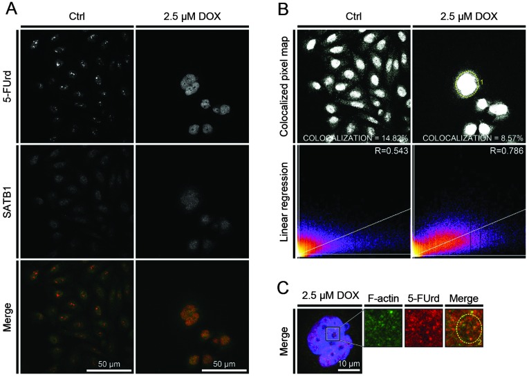
Colocalization of sequence-binding protein 1 (SATB1) and 5′-fluorouridine (5-FUrd). CHO AA8 cells were treated with 2.5 μM doxorubicin for 24 h, incubated with 5-FUrd for 20 min and labeled for both 5-FUrd and SATB1. (A) Localization of SATB1 and 5-FUrd in the control cells and cells treated with 2.5 μM doxorubicin. (B) Analysis of fluorescence colocalization of SATB1 and 5-FUrd using a confocal microscope in the control cells and cells treated with 2.5 μM doxorubicin. (C) Fluorescence colocalization of SATB1 and 5-FUrd in the area of nucleus of cells treated with 2.5 μM doxorubicin. Numbered dotted cirles indicate: 1, colocalization of 5-FUrd and SATB1 in the area of the nucleus of cells undergoing active cell death; 2, colocalization of 5-FUrd and SATB1 in the weak DNA labeling area of the nucleus of cells undergoing active cell death. Ctrl, control; DOX, doxorubicin.

