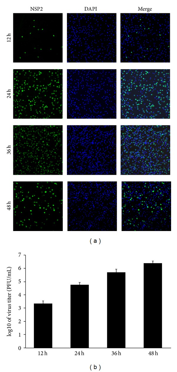
Figure 1.

Infection kinetics of the highly pathogenic PRRSV strain WUH3 in PAMs. (a) PAMs were infected with the adapted PRRSV (3rd passages) at an MOI of 0.1. Cells were fixed and permeabilized in cold methanol at different time point (12, 24, 36, and 48 h) postinfection. Immunofluorescence assays were performed to analyze the replication of PRRSV by detecting the nonstructural protein Nsp2 (green fluorescence). DAPI (4′,6-diamidino-2-phenylindole) was used to stain the nuclei. (b) PAMs were infected with the adapted PRRSV at a MOI of 0.1. Supernatants were collected at different time point (12, 24, 36, and 48 h) postinfection for plaque assay to determine viral titers.
