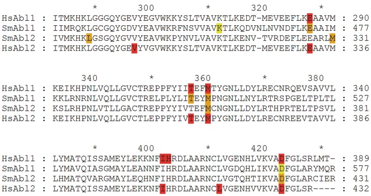
Figure 2. Alignment of the TK domains of human and schistosome Abl kinases.
Alignment: Structural alignment of the binding sites of human (HsAbl1, HsAbl2) and S. mansoni (SmAbl1, SmAbl2) Abl kinases focused on the highly conserved catalytic tyrosine domain [37]. Amino acid residues partnering in directional interactions with Imatinib are highlighted ocher and red for SmAbl1/2 and HsAbl1/2, respectively. Amino acids D568 and K457 of SmAbl1 are colored light teal, because they are not direct interaction partners to Imatinib but utilize a water molecule.

