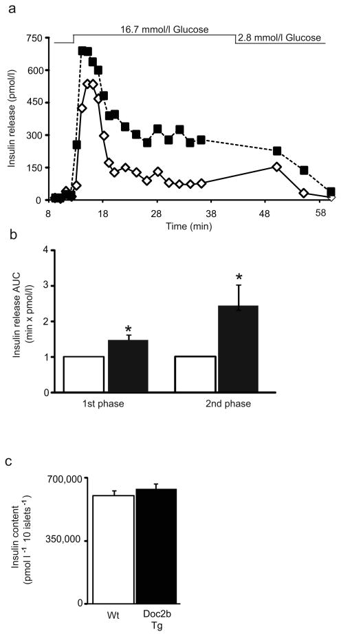
Fig. 4.
Islets from Doc2b Tg mice exhibit potentiated biphasic insulin release. (a) Islets isolated from Doc2b Tg mice (black squares) and Wt littermates (white diamonds) were perifused in parallel at 2.8 mmol/l glucose for 10 min followed by 16.7 mmol/l glucose for 35 min and then returned to low glucose for 20 min. Eluted fractions were collected and insulin secretion was determined by RIA, as depicted in this representative pair of traces. (b) AUC for first (11–17 min) and second (18–45 min) phases of insulin secretion was quantified in islets, normalised to baseline, from Doc2b Tg (black bars) and Wt (white bars) mice. Data represent the average ± SE from three independent sets of perifused islets; *p<0.05 vs Wt (Wt set equal to 1.0 and Tg normalised thereto for each phase per set). (c) Average insulin content per 10 islets from Doc2b Tg mice and Wt littermates

