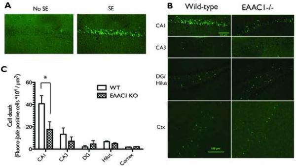
Figure 3. Photomicrograph of representative Fluoro-Jade C staining from wild-type and EAAC1−/− animals 48 hours after pilocarpine-induced seizures and comparison of cell death.
(A) Fluoro-Jade staining is evident in the hippocampi of mice that have experienced SE, whereas it is absent in the hippocampi of mice that received pilocarpine but did not experience SE. CA1 pyramidal cells in wild-type animals were more sensitive to pilocarpine-induced cell death than those in CA3, the dentate gyrus, the hilus, or the cortex. (B) In EAAC1−/− mice, less cell death was consistently observed in CA1 than was observed in the wild-type mice. (C) A two-way ANOVA shows a significant effect of genotype on the amount of cell death following pilocarpine-induced SE, which a post hoc test shows to be due to a significant difference in CA1. EAAC1−/− mice had significantly less cell death in the CA1 region of the hippocampus than wild-type animals (WT = 40.77 ± 7.27 * 104 cells/μm2, KO = 17.76 ± 6.80 * 104 cells/μm2, p = 0.03). Data are presented as mean ± SEM, WT n=7, KO n = 10 animals.

