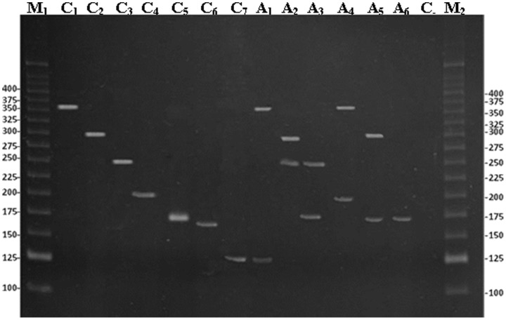
Figure 1. Electrophoretic analysis of the amplified fragments by using a multiplex polymerase chain reaction in 8% polyacrylamide gel stained with ethidium bromide.
Lane C1: control of Chlamydia trachomatis (361 base pairs-bp); lane C2: control of Treponema pallidum (291 bp); lane C3: control of HSV–2 (249 bp); lane C4: control of Mycoplasma genitalium (193 bp); lane C5: control of Trichomonas vaginalis (170 bp); lane C6: control of Neisseria gonorrhoeae (162 bp); lane C7: control of HSV–1(123 bp); lane A1: positive sample of C. trachomatis and HSV–1 (361 and 123 bp); lane A2: positive sample of T. pallidum and HSV–2 (291 and 249 bp); lane A3: positive sample of T. vaginalis and HSV–2 (170 and 249 bp); lane A4: positive sample of C. trachomatis and M. genitalium (361 and 193 bp); lane A5: positive sample of T. pallidum and T. vaginalis (291 and 170 bp); lane A6: positive sample of T. vaginalis (170 bp); lanes M1 and M2, molecular weight marker (25 bp Invitrogen). Values on the left and right sides of the gel are in bp.

