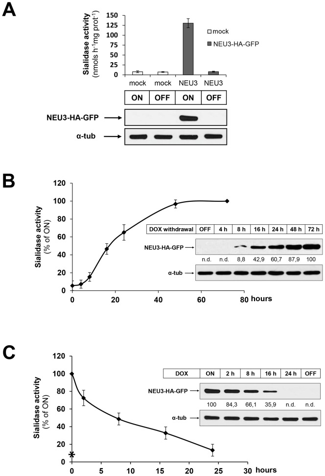
Figure 1. Characterization of the inducible expression cell model HeLa tTA2 NEU3-HA-GFP.
(A) HeLa tTA2 cells stably transfected with pUHD 10.3 NEU3-HA-GFP or with the empty vector (mock) were grown for 7 days in absence (ON) or presence (OFF) of dox. Cell homogenates were analyzed for sialidase activity (values are given as mean +/− SD and represent the mean of 5 independent experiments) and for NEU3-HA-GFP expression using anti-HA primary antibody. Alpha-tubulin was detected with specific primary antibody in order to normalize NEU3-HA-GFP signal. (B) OFF HeLa tTA2 NEU3-HA-GFP were plated and grown in absence of dox for the indicated time periods. Cell homogenates were analyzed for sialidase activity (values refer to ON cells and represent the mean +/− SD of 4 independent experiments) and NEU3-HA-GFP expression using anti-HA primary antibody. NEU3-HA-GFP optical density was normalized to alpha-tubulin as indicated in (A) and values are given. (C) ON HeLa tTA2 NEU3-HA-GFP were plated and grown in presence of dox for the indicated time periods. Cell homogenates were analyzed for sialidase activity and NEU3-HA-GFP expression as indicated in (B). Asterisk indicates the sialidase activity measured in OFF cells.

