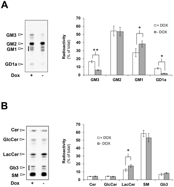
Figure 2. Expression of NEU3 results in the modification of the cellular ganglioside composition.
OFF HeLa tTA2 NEU3-HA-GFP cells were plated and further grown for 72 h in absence (ON) or presence (OFF) of dox. Cells were then metabolically labeled for 2 h with [3H]-sphigosine and chased for 48 h. Cell lipids were extracted and gangliosides (A) and non-ganglioside sphingolipids (B) were separated by HPTLC and visualized (left, Beta-Imager 2000) and associated radioactivity was determined (right, Beta-Vison). Values are given as percentage of total radioactivity and represent the means ± S.D. of 5 independent experiments. *p<0.05; **p<0.01.

