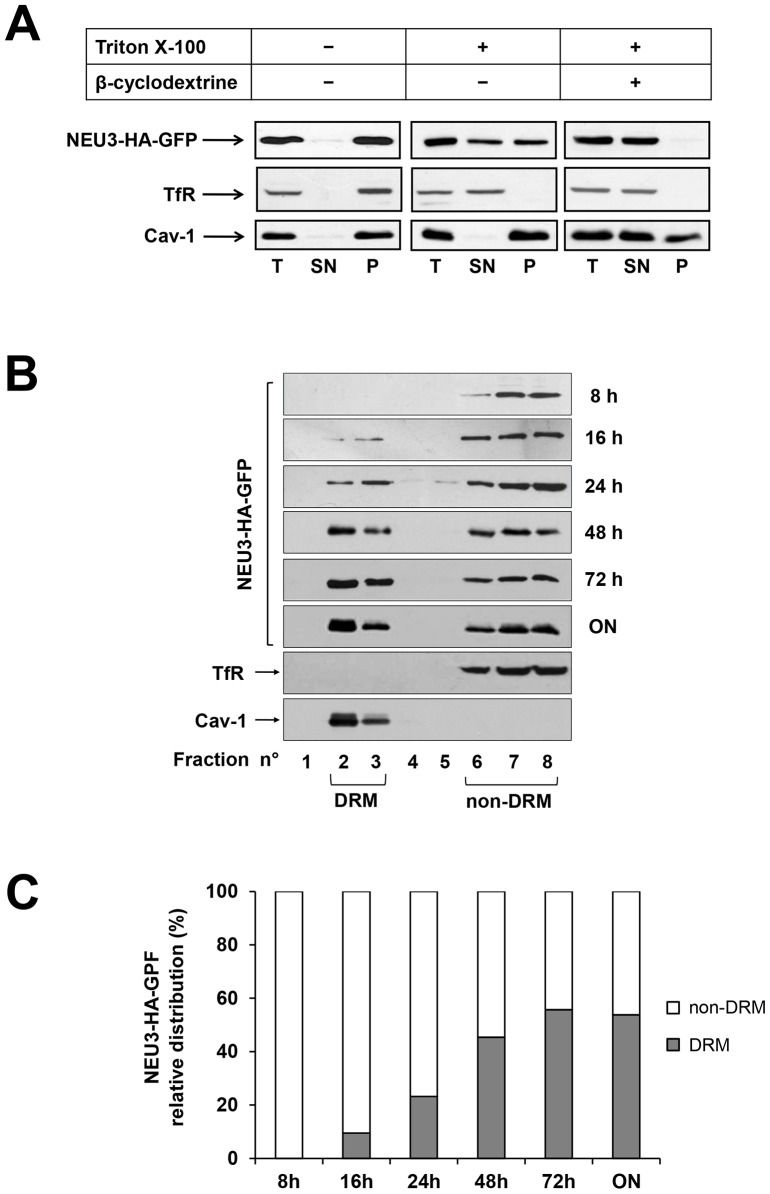
Figure 3. Association of NEU3-HA-GFP to DRM and non-DRM.
(A) ON HeLa tTA2 NEU3-HA-GFP grown for 1 h in absence or presence of beta-cyclodextrine were extracted in the appropriate buffer, containing or not 1% Triton X-100 for 30 min at 4°C and ultracentrifuged for 1 h at 100,000×g, Equal amounts of total (T), supernatant (SN) and pelleted (P) material was analyzed by western blot using anti-HA, anti-Transferrin Receptor (TfR) and anti-caveolin-1 (Cav-1) primary antibodies. (B) OFF HeLa tTA2 NEU3-HA-GFP were grown in absence of dox for the indicated time periods and extracted in the appropriate buffer containing 1% Triton X-100 for 30 min at 4°C. DRM and non-DRM were separated by Opti-Prep density gradient centrifugation. Equal amounts of each gradient fraction were analyzed by western blot as indicated in (A). (C) Relative distribution of NEU3-HA-GFP between DRM (fractions 2 and 3) and non-DRM (fractions 6 to 8) referred to the optical density of bands in (B).

