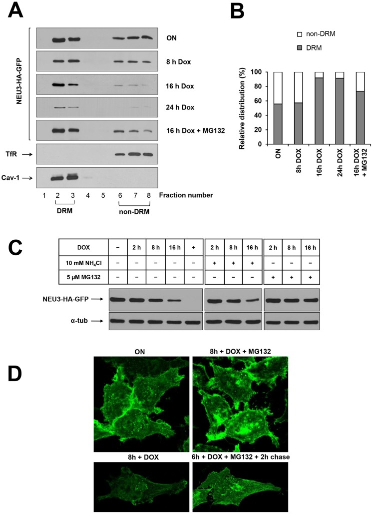
Figure 5. NEU3-HA-GFP is specifically degraded by the proteasomal machinery.
(A) ON HeLa tTA2 NEU3-HA-GFP were grown in presence of dox for the indicated time periods, with or without 5 µM MG132, and extracted in the appropriate buffer containing 1% Triton X-100 for 30 min at 4°C. DRM and non-DRM were separated by Opti-Prep density gradient centrifugation. Equal amounts of each gradient fraction were analyzed by western blot using anti-HA, anti-Transferrin Receptor (TfR) and anti-Caveolin-1 (Cav-1) primary antibodies. (B) Relative distribution of NEU3-HA-GFP between DRM and non-DRM referred to the optical density of bands in (A). (C) ON HeLa tTA2 NEU3-HA-GFP were grown in presence of dox for the indicated time periods, with or without NH4Cl or MG132. NEU3-HA-GFP expression was analyzed by western blot using anti-HA primary antibody. Alpha-tubulin was detected with specific primary antibody and used as control for total protein loaded on gel. (D) ON HeLa tTA2 NEU3-HA-GFP were plated onto glass coverslips and grown in presence of dox for 8 h, with or without MG132. In order to test reversibility effect of MG132, one sample was incubated for 6 h with MG132, followed by 2 h chase in normal growth medium. After fixation, GFP signal was detected by laser scanning microscope using the same settings (laser power, gain and offset) for all images.

