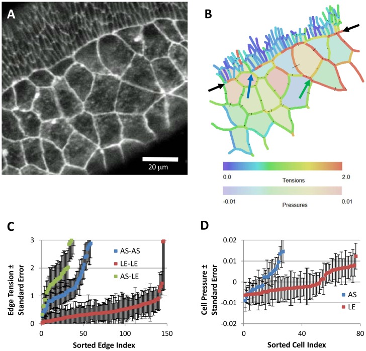
Figure 5. CellFIT analysis of cells near the amnioserosa/lateral epidermis boundary during early dorsal closure in a living Drosophila embryo, as imaged in (A) and with inferred Standard Tensions and Pressures illustrated in (B) according to the color bars.
The amnioserosa is visible in a wide band from the lower left-hand corner towards the upper right, while the lateral epidermis, identifiable by its smaller cells, is confined to a large triangle in the upper left corner. The boundary between these two tissues is indicated by the black arrows. The blue and green arrows point to features discussed in the text. Overall, the CellFIT equations were very well conditioned – having tension and pressure condition numbers of 30.3 and 15.6, respectively. The tension and pressure residuals are shown in (B) as thin lines emanating from each triple junction and bisecting each cell edge, respectively. These residuals are scaled so that a residual equal to the mean tension has a length equal to the mean cell radius. Even at this scaling, the residuals are generally quite small and many are barely discernable. Finally, confidence limits are shown for individual tensions and pressures, (C) and (D) respectively, by boundary type. The points and bars indicate best estimate +/− one standard error (based on the covariance matrix), respectively. These confidence limits are a significant, but modest, fraction of the inferred tensions and pressures. Prior investigations have suggested the existence of a uniform high-tension purse-string along the edge of the amnioserosa, but CellFIT reveals a more complex and interesting scenario. See text for details.

