
Figure 1.
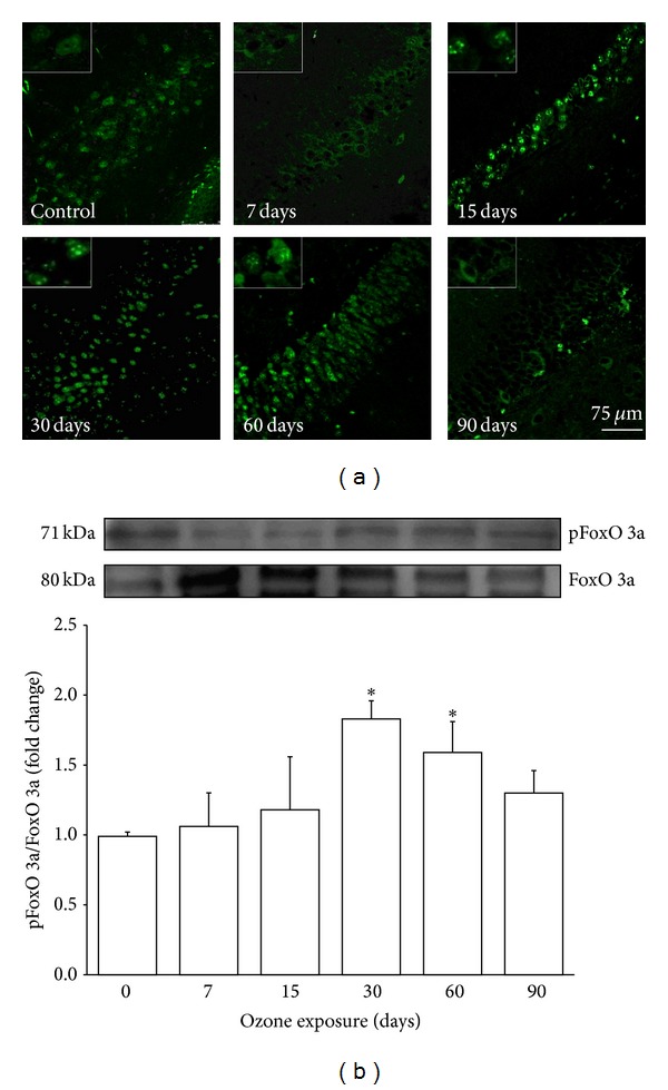
Effect of ozone exposure on pFoxO 3a in the rat hippocampus. (a) The micrographs show cells that are positive for phosphorylated FoxO 3a (pFoxO 3a) (green) in the dentate gyrus (DG) of rats exposed to ozone-free air (control) or 7, 15, 30, 60, and 90 days of ozone. The images show a progressive increase in the immunoreactivity against phosphorylated FoxO 3a from 15 days to 60 days of exposure to ozone. pFoxO 3a shows nuclear localization from 15 days to 60 days of ozone exposure and at 90 days is located in the cytoplasm. Insets are a zoom from selected areas in the same picture. (b) A representative western blot shows the contents of total FoxO 3a and pFoxO 3a in the homogenated hippocampi of rats exposed to ozone for different durations. The normalized graph shows densitometry values presented as the mean ± SD (∗ P < 0.05) (n = 6).