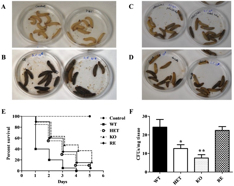
Figure 8. Virulence of C. parapsilosis.
The degree of melanization in response to fungal infection was monitored by visual inspection of the G. mellonella larvae in control (A), WT strain (B), KO strain (C) and the reintegrant (RE) C. parapsilosis strain (D). Survival of G. mellonella larvae infected with 2.5×10⁶ yeast cells from each strain in PBS was monitored over time (E). The ability of C. parapsilosis to invade tissues was evaluated in a mouse model after intraperitoneal infection of Swiss mice (F). After 3 days of infection, the wild type (WT), heterozygous (HET), homozygous (KO) and reintegrant (RE) yeast strains were recovered from the kidneys and the CFUs/mg tissue were calculated. Ten mice were used per group and the experiment was repeated twice with similar results. Student t-test: *P<0.05; **P<0.01 between WT and HET, KO or RE strains.

