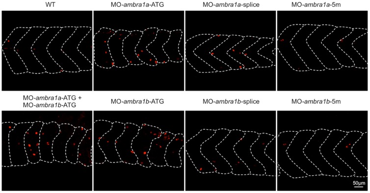
Figure 5. Cell proliferation in muscles of 3 dpf control and ambra1 morphant embryos.
Mitotic cells, detected by immunostaining for phospho-histone H3 in longitudinal sections, are more abundant in ATG-morphant embryos with respect to WT and 5 m-control embryos. Anterior is to the left and dorsal up.

