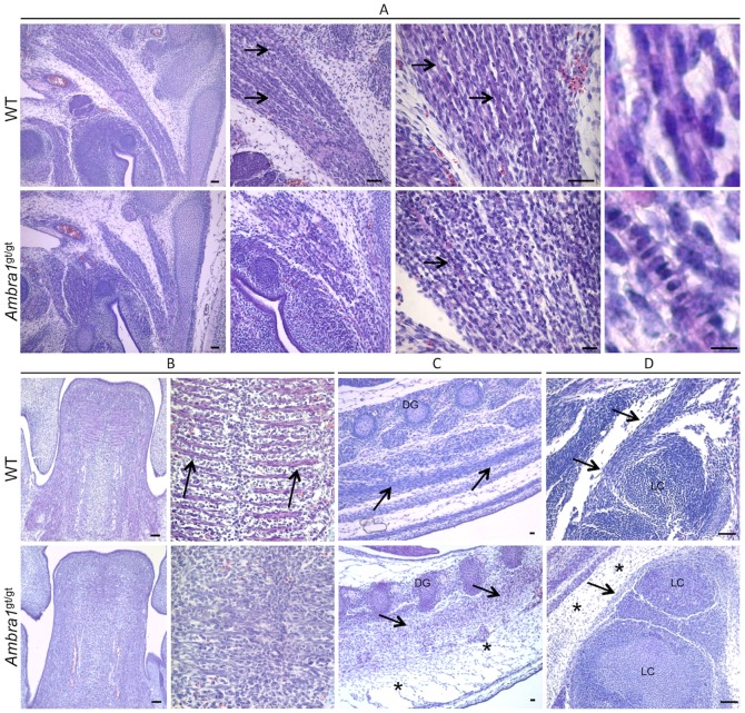
Figure 10. Morphological alteration of skeletal muscles in Ambragt/gt mouse embryos.
Representative pictures of skeletal muscles from WT and Ambra1 gt/gt E13.5 mouse embryos, following haematoxylin-eosin staining. (A) Details of neck muscle. WT embryos display several well-organized and mature myofibers (black arrows), which have myonuclei already localized at the edge of the cell (right panel). In Ambra1 gt/gt embryos, the muscle is much more immature, with poorly organized myofibers displaying centrally located nuclei (black arrows). (B) Details of the tongue. In WT embryos, myofiber are formed and well-organized (arrows), whereas in Ambra1 gt/gt embryos there is a general disorganization of muscle architecture. (C, D) Representative pictures of dorsal (C) and limb (D) muscles (black arrows). Ambra1 gt/gt embryos show abnormal muscle organization, together with a marked thickening of the connective tissue (black asterisks). All the analyzed muscles also display a noticeable increase of cell density in Ambra1 gt/gt embryos. Scale bar, 50 µm.

