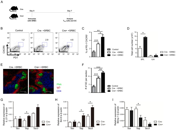
Figure 7. PPARγ deficiency in T cells increases follicular helper T cell and germinal center formation following SRBC immunization.
(A) Experimental scheme for SRBC immunization to analyze TFH cells and GC reaction. (B, C) Flow cytometric analysis of TFH cells in the spleen and (D) total cell number of spleens and lymph nodes from 8-week-old control and SRBC-immunized mice. Values represent the mean ± SEM, n = 7–10. *P<0.05, **P<0.01, ***P<0.001. (E) Germinal centers of frozen 7-µm sections from spleen of 8-week-old control (non-immunized Cre-) and SRBC-immunized mice were visualized by confocal microscopy. Slides were stained for PNA (green), IgD (red), and CD4 (blue) to detect germinal centers, B cells, and T cells, respectively. (F) The number of PNA+ germinal centers per spleen section was quantitated n = 5–7. Expression level of (G) IL-21, (H) Bcl-6 and (I) Blimp-1 was assessed by real-time PCR and normalized to β-actin. Values represent the mean ± SEM, n = 3. **P<0.01, ***P<0.001.

