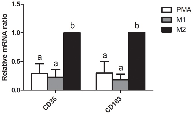
Figure 1. Expression of markers for M2-type macrophages in phorbol 12-myristate 13-acetate (PMA)-treated THP-1 macrophages. THP-1 cells treated with 320 nM PMA for 4 h, and then cultured with PMA plus 20 ng/mL interleukin (IL)-4 and 20 ng/mL IL-13 for another 20 h had significantly higher mRNA expressions of CD36 and CD163 (both markers of M2 macrophages) compared to those of M1-polarized THP-1 macrophages.

mRNA expression was analyzed by a real-time PCR. Multiples of mRNA changes were quantitated by the comparative CT method. All data are presented as the mean ± SD, n = 6. Means with different letters significantly differ (p<0.05).
