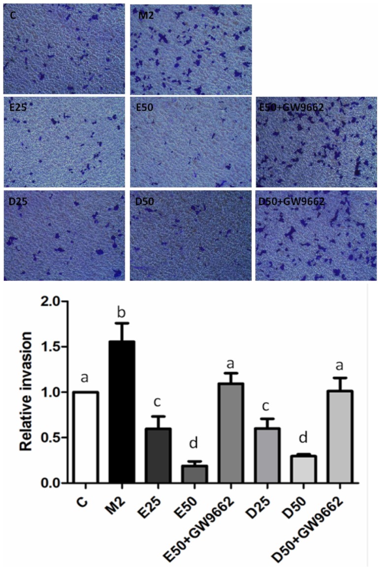
Figure 5. Effects of EPA and DHA on PC-3 prostate cancer cell M2-type macrophage-induced invasion.

For the invasion assay, PC-3 cells were seeded in the upper chambers coated with Matrigel and cocultured with M2-polarized THP-1 macrophages in the presence of 50 µM EPA, DHA, or GW9662 (10 µM) for 24 h. Invaded cells were fixed in 4% paraformaldehyde and stained with 0.5% crystal violet. Membranes were washed, and the dye was eluted with a violet extraction solution (50% ethanol, 0.1% acetic acid, and 49.9% ddH2O). The absorbance was measured at 595 nm using a microtiter plate reader. Data are presented as the mean ± SD, n = 3. Means with different letters significantly differ (p<0.05).
