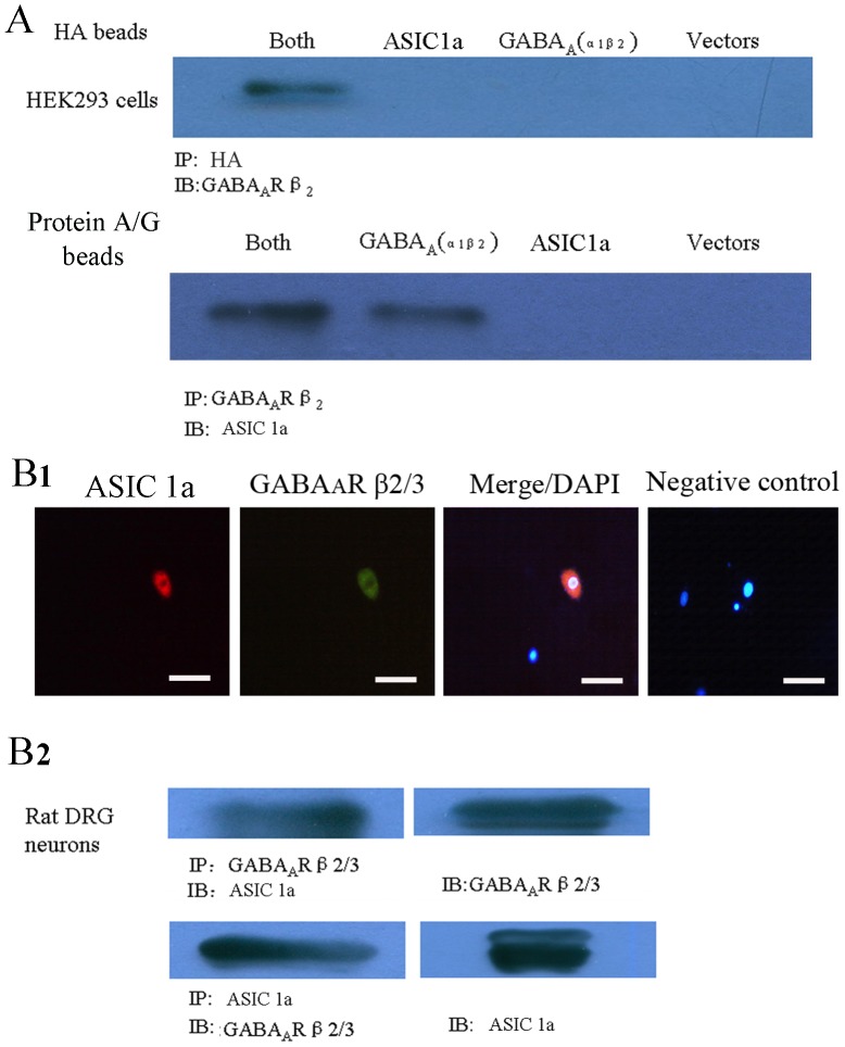
Figure 4. Co-immunoprecipitation of ASIC1a and GABAA proteins in transfected HEK293 cells and DRG neurons.
A. Co-immunoprecipitation of ASIC1a and GABAA proteins in transfected HEK293 cells. GABAA specifically co-precipitates with ASIC1a in cells co-transfected with ASIC1a and GABAA (both). n = 3. B1. DRG neurons were double-labeled with anti-ASIC1a and -GABAAR antibodies. ASIC1a (red in left panel) as well as GABAAR (green in middle panel) localized to the apical membrane of DRG neurons. The merge (right) of ASIC1a and GABAAR images indicates that ASIC1a co-segregates with GABAAR in the apical membrane in neurons (yellow). Nuclei were identified by DAPI staining (blue). Scale bars equal 50 µm. B2. GABAA precipitates with ASIC 1a in primary cultured DRG neurons. DRG neurons lysates were immunoprecipitated (IP) with anti-GABAA β2/3 subunits polyclonal antibody in 8% SDS-PAGE gel, and the blot was then probed with anti- ASIC 1a polyclonal antibody (IB). In turn, ASIC 1a co-immunoprecipitates with GABAA. Cell lysates were immunoprecipitated with ASIC 1a antibody in 6% SDS-PAGE gel, and the blot was probed with GABAA β2/3 antibody. These experiments were repeated three times with identical results.

