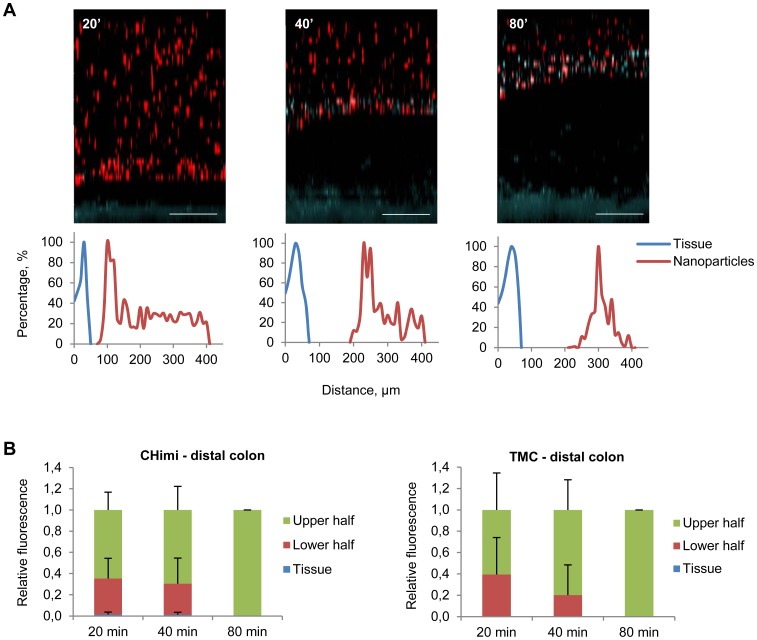
Figure 5. Mucus penetrability of nanoparticles in mouse distal colon explants.
(A) Representative Z – stack projections of TMC/siRNA nanoparticles in distal colon explants and the corresponding normalised intensity plots; tissue is blue and nanoparticles are red. Scale bars 100 µm. (B) Percentage of the total fluorescence intensity of TMC and CHimi2/siRNA nanoparticles in each plan (tissue, lower half, and upper half) at each time point. Data are presented as means ± SD (n = 3).

