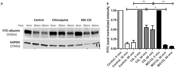
Figure 2. Albumin degradation in podocytes decreased with lysosome inhibition, but not proteasome inhibition.
Densitometry and representative Western blot demonstrating albumin degradation. Podocytes were pretreated with standard media (controls, n = 7), 50 µM chloroquine (lysosome inhibitor, n = 5), or 10 µM MG-132 (proteasome inhibitor, n = 4) for 24 h. At 24 h, podocytes were treated with 1.5 mg/mL FITC-albumin for 1 h then washed. Cells were harvested at time zero, 30 min, and 60 min after washing. A) Albumin abundance in cell lysates was quantified by Western blot analysis using an antibody to FITC. B) Albumin abundance in chloroquine treated cells was significantly increased compared to control and MG-132 treated cells at 30 min (*p<0.05) and 60 min (**p<0.05). Densitometry was normalized to GAPDH and time zero.

