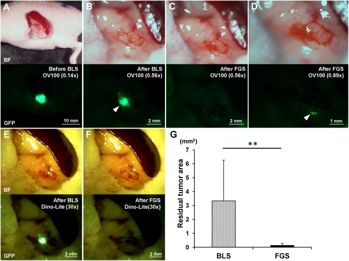
Figure 2. Preoperative and postoperative images of the orthotopic pancreatic cancer model (A–F).
Upper panels are bright-field (BF), and lower panels show tumor fluorescence. The residual tumor after BLS was clearly detected with both the OV100 at a magnification of 0.56x (B) and the Dino-Lite at a magnification of 30x (E). The residual tumor after FGS was marginally detected with either the OV100 at a magnification of 0.56x (C) or the Dino-Lite at a magnification of 30x (F). The OV100 at a magnification of 0.89x clearly detected the minimal residual tumor after FGS (D). (G) The residual tumor area after FGS was significantly smaller than after BLS. All images were measured for residual tumor areas using ImageJ. **p<0.01.

