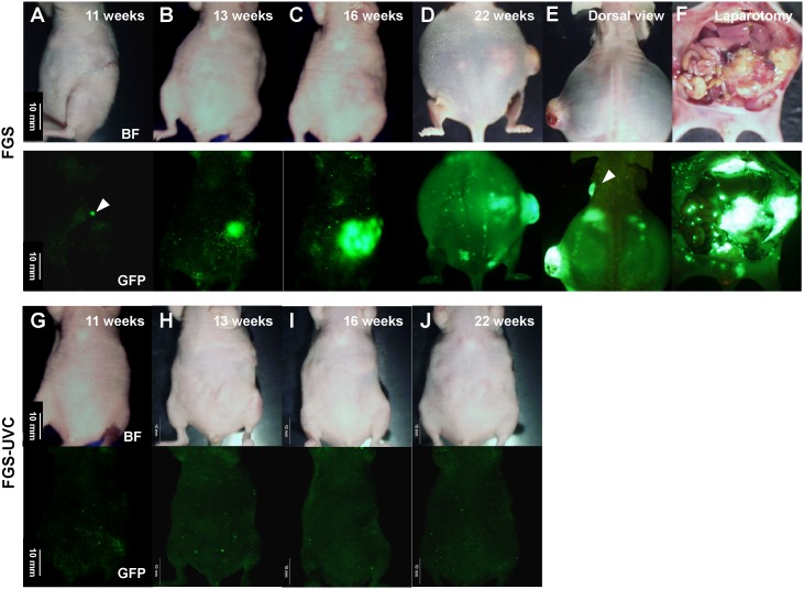
Figure 3. Representative time course of tumor recurrence after FGS.
The recurrence was initially detected by non-invasive whole-body imaging using the OV100 at a magnification of 0.14x at week 11 after FGS (A; white arrowhead). The recurrent tumor progressed rapidly (B–D) and killed the mice by week 22 after FGS (D). Left axillary lymph-node metastasis (E; white arrowhead), large local recurrent tumor and many disseminating tumor nodules (F) were detected in the mice at time of death. In contrast, no recurrence was detected in the FGS-UVC group (G–J). Scale bars: 10 mm.

