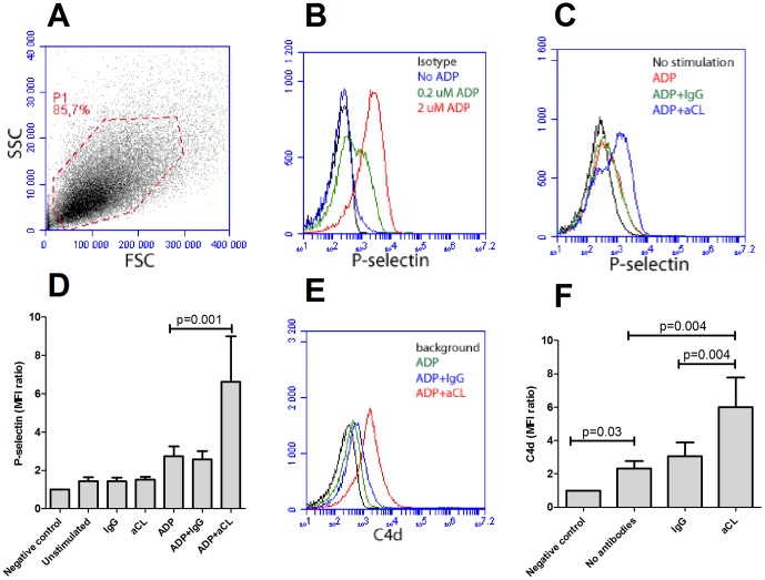
Figure 1. Anti-phospholipid (aPL) antibody-mediated platelet activation and complement deposition.
A) Isolated platelets from healthy individuals were gated by flow cytometry (P1) and had consistently purity above 98%. B) Platelet activation after ADP stimulation was detected as P-selectin expression. The lower concentration (0.2 µM) was used for sub-optimal activation. C) Representative figure of P-selectin expression in sub-optimal activated platelets with or without addition of anti-cardiolipin antibodies (aCL). D) A summary of the data presented in Figure 1C. The results are the mean and standard deviation of six or more independent experiments. E–F) Activated and fixed platelets were incubated with or without aCL antibodies in presence of normal human serum. Complement deposition was analyzed with flow cytometry and illustrated as E) a representative histogram and F) a summary of the mean and standard deviation of six or more independent experiments.

