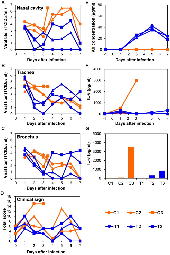
Figure 3. Protection of immunocompetent macaques treated with MAb ch61 against VN3040 infection.
Macaques infected with VN3040 (3×10⁶ PFU) on day 0 were injected intravenously with control MAbs (C1–C3, orange) or MAb ch61 (T1–T3, blue) on days 1 and 3. Viral titers in nasal (A), tracheal (B), and bronchial (inside lungs) (C) swab samples were determined using MDCK cells. Viral titers under the detection limit are indicated as 0. Clinical signs were scored with the parameters shown in Table S1 (D). Serum samples were collected from macaques during the period of the experiments and antibodies specific to influenza virus HA were quantified by ELISA [23] (E). IL-6 concentrations in serum samples (F) and lung tissue samples (G) were measured as described in the Materials and Methods section. Lung tissue samples of macaque C3 and other macaques were collected at autopsy on day 3 and on day 7, respectively, and IL-6 concentrations in 10% (w/v) homogenates in saline were measured.

