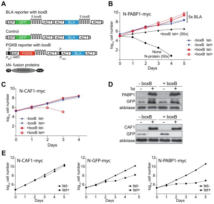
Figure 1. Proof of concept of the tethering approach.
(A) Schematic diagram of reporter mRNAs and tethered proteins, not to scale. Above: BLA and GFP reporter mRNA with five boxB recognition sites in the 3′-UTR. Middle: Control BLA 3′-UTR mRNA without boxB. Below: PGKB mRNA with boxB recognition sites at the 3′-UTR. A tetracycline-inducible EP1 procyclin promoter (flag) drives PGKB expression. The downstream promoter, PVSG, gives constitutive expression of the resistance marker. Bottom: Fusion protein with N-terminal lambda-N peptide and C-terminal myc tag. (B) Cumulative growth curves of cells expressing the BLA reporters with tetracycline-inducible lambda-N-PABP1, in the presence of 5x blasticidin (25 µg/ml) with or without tetracycline. For comparison, cells expressing lambda-N-PABP1 and non-transfected cells (without N-protein, parental cell line) grown in 50x blasticidin (250 µg/ml) are shown. The +boxB cells with lambda-N-PABP express a little of the lambda-N fusion even in the absence of tetracycline (panel D), which may result in slightly better growth in the presence of 25 µg/ml blasticidin. (C) Cumulative growth of cells containing the BLA-boxB reporter expressing N-CAF1-myc in the presence or absence of tetracycline and 5x blasticidin. (D) Western blot analysis of N-PABP1-myc, N-CAF1-myc and N-GFP proteins. Expression of PABP1-myc and CAF1-myc was detected with anti-myc, using aldolase as loading control. (E) Effect of tethered PABP1, GFP and CAF1 on growth after tetracycline-mediated induction of both PGKB and tethered protein expression.

