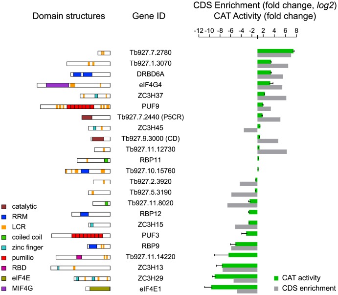
Figure 5. Validation of mRNA-fate regulators.
Expression of CAT in cells expressing different myc-tagged lambda-N-fusion proteins was assayed after 24 h tetracycline induction and activities were expressed relative to a control with no lambda-N protein (green). Results are arithmetic mean ± standard deviation of at least 3 independent clones. As comparison, the fold per CDS (log2 values) are shown (grey). (left) Domain structures of analysed T. brucei proteins as detected by SMART (http://smart.embl-heidelberg.de). Different domains are specific colours as shown on the blocks. P5CR, pyrroline-5-carboxylate reductase; CD, cytidine deaminase; LCR, low-complexity region; RRM, RNA recognition motif; RBD, RNA-binding domain. RBP11 and Tb927.10.15760 are negative controls, which did not have reproducible effects in the tethering screen.

