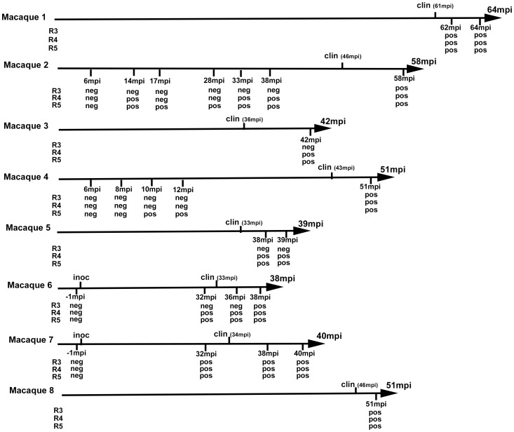
Figure 5. vCJD agent detection in the buffy coat of experimentally infected primates.
Eight cynomologus macaques were intravenously challenged with vCJD brain homogenate or blood from a vCJD affected macaque (macaque 6). At different time points of the incubation period, blood was collected and buffy coat prepared. Clinical onset (clin) and time to euthanasia of the animals are indicated (upper label on arrows) as months post inoculation (mpi). The buffy coat samples were used (as homogenates 1/100 diluted in PMCA buffer) to seed PMCA reactions in which brain homogenate from ovine PrP transgenic mouse (ARQ variant) was used as substrate. Each sample was submitted to 6 rounds of amplification each comprising 96 cycles (30 s sonication-30 minutes incubation at 39.5°C) in a Misonix 4000 sonicator. PMCA products were analyzed by Western Blot (WB) for the presence of abnormal PK resistant PrP (PrPres -antibody Sha31 epitope YEDRYYRE). Samples were received encoded and tested blind. The time point corresponding to blood samples (months post inoculation) that were tested and the results of PrPres WB detection in PMCA reactions are indicated (under arrow). No positive WB result was observed before the third PMCA round. No additional positive result was observed after 5 PMCA rounds.

