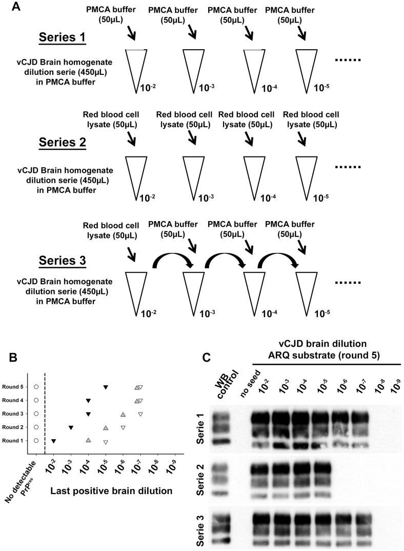
Figure 7. Red blood cell and inhibition of PMCA vCJD amplification.
(A) Two vCJD brain homogenate (human) dilution series (1/10 dilution, 10−2 to 10−9) were prepared (aliquots of 450 µL). Each aliquot also contained 50 µL of PMCA buffer (series 1: ▿) or Red blood cell lysate (human) (series 2:▾). A third series (series 3:  ) was prepared starting from a 10−2 dilution of the vCJD brain homogenate (900 µL) in which 100 µL of red blood cell lysate have been added. In each aliquot (450 µL) of dilution series 3, 50 µL of PMCA buffer were added. The three dilution series were then used to seed PMCA reactions (7 µL of seed) in which ovine PrP expressing mice (ARQ variant) was used as substrate (63 µL). Five successive rounds of PMCA were performed. After each round PrPres detection was carried out in PMCA reactions by Western blot (Sha31 anti PrP monoclonal antibody: epitope: YEDRYYRE, amino acid 145–152). Ten unseeded controls (○) were included in the experiment. The results of the PMCA amplification after each round are presented in graph (B). WB corresponding to the fifth round of amplification is presented as an illustration (C).

