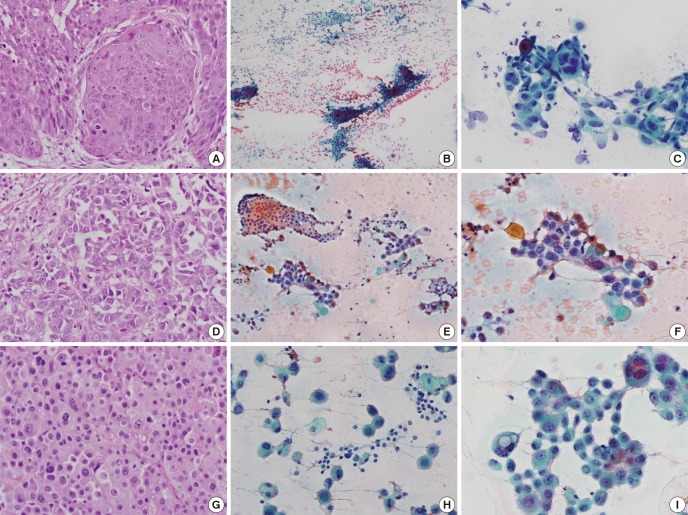
Fig. 2.
The histology shows the primary foci and cytological features of matched thyroid lesions (A-C, Case 2; D-F, Case 11; G-I, Case 13). (A) Tumor nests of a well-differentiated squamous cell carcinoma are observed in a specimen of right bronchus. (B, C) Thyroid cytology of Case 2 reveals a slight inflammatory background, and two populations of epithelial cells: follicular epithelial cells and tumor cells. Dyskeratotic cells are also observed in the tumor clusters. (D) A moderately differentiated adenocarcinoma is seen, with frequent mitotic figures. (E, F) The cytology of Case 11 shows a colloid background and heterogeneous population. (G) A poorly differentiated adenocarcinoma is seen. (H, I) Smear of the thyroid aspirate shows large hyperchromatic atypical cells with occasional intracytoplasmic vacuoles. (A, D, G, hematoxylin and eosin, ×400; B, C, E, F, H, I, Papanicolaou stain, ×400).

