
Fig. 3.
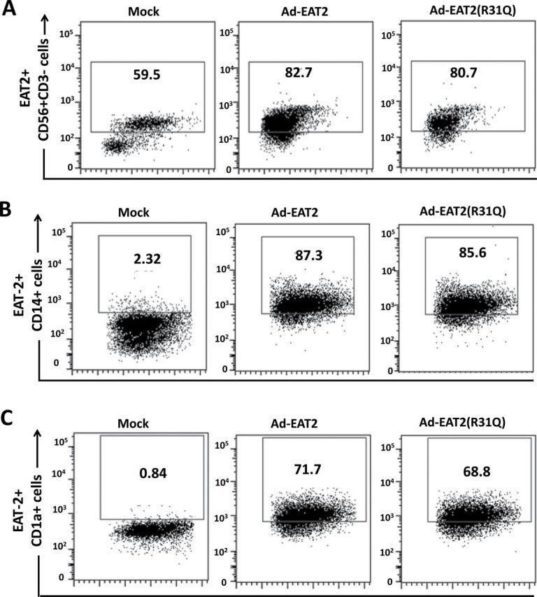
Intracellular staining analysis for EAT-2 expression in human immune cells following rAd5-EAT2 infection. Human PBMCs were isolated from fresh buffy coat material and were either mock infected or infected with MOI of 10000 with rAd5-EAT2 or rAd5-EAT2(R31Q). Intracellular staining analysis for EAT-2 expression at 48h following Ad infection of human NK cells (A), CD14+ monocytes (B) and CD1a+ DCs (C) is shown. Data were collected using a BD LSRII flow cytometer and analyzed using FlowJo software.