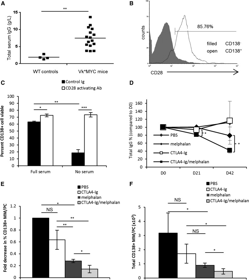
Figure 2.
CD28 signaling protects Vk*MYC murine myeloma cells in vitro and blockade sensitizes MM to melphalan in vivo. (A) Seventy-two-week-old Vk*MYC mice or their wild-type (WT) littermate controls were screened by ELISA for total serum IgG levels. (B) Whole bone marrow from Vk*MYC mice was isolated and the percent of myeloma cells was determined using a CD138+CD28+B220−CD38−MHCII−CD3− phenotype. (C) Purified CD138+ cells from disease-bearing Vk*MYC mice were plated for 24 hours in media containing 10% fetal bovine serum (full serum) or no serum. Cells were cultured with either hamster Ig-coated beads (isotype control) or CD28-activating antibody-coated beads (clone PV1) at a ratio of 2 beads:1 cell. Cell viability was assessed by trypan blue exclusion. (D) Based on titers as in panel A, mice were randomized into 4 treatment groups (n = 3-4 mice/group): PBS, melphalan alone (2.0 mg/kg), CTLA4-Ig alone (100 μg/mouse), or melphalan plus CTLA4-Ig. Mice were treated intraperitoneally every third day for 42 days and serum samples were drawn weekly. Total serum IgG was determined by ELISA and was plotted as total IgG percent compared with day 0. (E) Percent MM/PC was determined using multiparametric flow for CD138+CD28+B220−CD38−MHCII−CD3− cells, and fold decrease was calculated compared with the PBS group. (F) Total numbers of MM/PC were calculated by multiplying the percent MM/PC as determined in panel E by the total number of cells counted for each tissue. Data are representative of 2 separate experiments. *P < .05, **P < .01, ***P < .005.

