Figure 4.
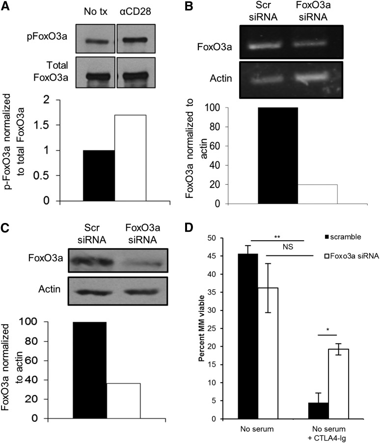
CD28 signaling induces phosphorylation of FoxO3a and knockdown of FoxO3a partially prevents CD28 blockade–induced apoptosis. (A) MM.1S cells were cultured in serum-free media for 24 hours ± 10 μg/mL αCD28.2. Cells were then analyzed by western blot for phospho-FoxO3a (top) or total FoxO3a (bottom). Densitometry was assessed using Quantity One software. (B) MM.1S cells were cultured for 24 hours in melphalan ± 10 µg/mL αCD28.2. Cells were lysed and RNA was collected. Semiquantitative RT-PCR was conducted (top) and assessed by densitometry (bottom). (C) A total of 1 × 10⁶ cells were transfected with FoxO3a or scramble siRNA and FoxO3a expression was assessed by western blot after 48 hours. Densitometry was assessed using Quantity One software and is compared with the scramble siRNA. (D) Cells were transfected with siRNA as in panels B-C; 48 hours later, cells were plated in serum-free medium ± 100 µg/mL CTLA4-Ig. Cells were harvested after 48 hours and survival was assessed by Annexin V/7AAD staining by flow cytometry. Data are representative of 3 separate experiments, except for panel B, which is representative of 2 separate experiments. *P < .05, **P < .01. tx, treatment.

