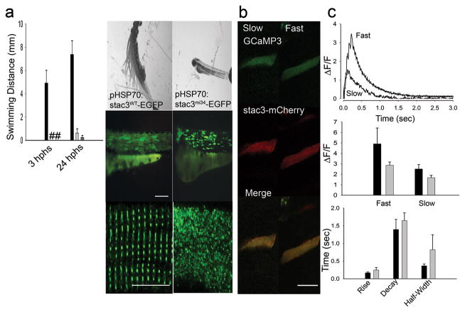
Fig. 4. Expression of wt Stac3 in Muscles Rescues the stac3mi34 Phenotype.
(a) Left, histogram showing that mutant embryos expressing heat inducible stac3wt-egfp (black, n=32) but not stac3mi34-egfp (light gray, n=4) nor uninjected mutant embryos (dark gray, n=10) exhibited touch evoked swimming at both 3 h post heat shock (hphs) and 24 hphs. # denotes no swimming. Right panels, superimposed frames (30 Hz, 1 sec) (top) show that mutant embryos expressing stac3wt but not stac3mi34 swim in response to touch although both are similarly expressed by muscles (middle, scale: 180 μm). In dissociated mutant muscles Stac3wt-EGFP localizes to the triads but not Stac3mi34-EGFP (bottom, scale: 10 μm). (b) In vivo expression of Stac3wt-mCherry by mutant muscle fibers rescues Ca2+ transients. Left panels, stac3mi34 mutant slow and fast twitch fibers co-expressing α-actin driven GCaMP3 and heat induced Stac3wt-mCherry. The triadic localization of Stac3wt-mCherry cannot be seen due to the low resolution of the resonance scans used to detect the fluorescent proteins. (c) Top, Ca2+ transients from mutant fast and slow fibers co-expressing Stac3wt-mCherry and GCaMP3. Middle, histogram showing that peak Ca2+ release is comparable between wt (black) fast (n=5) and slow (n=5) fibers and rescued mutant (gray) fast (n=6) and slow (n=2) fibers. Bottom, histogram showing that the kinetics of Ca2+ transients from wt (black) and rescued mutant (gray) fast twitch fibers are comparable. Error bars represent standard error of the means.

