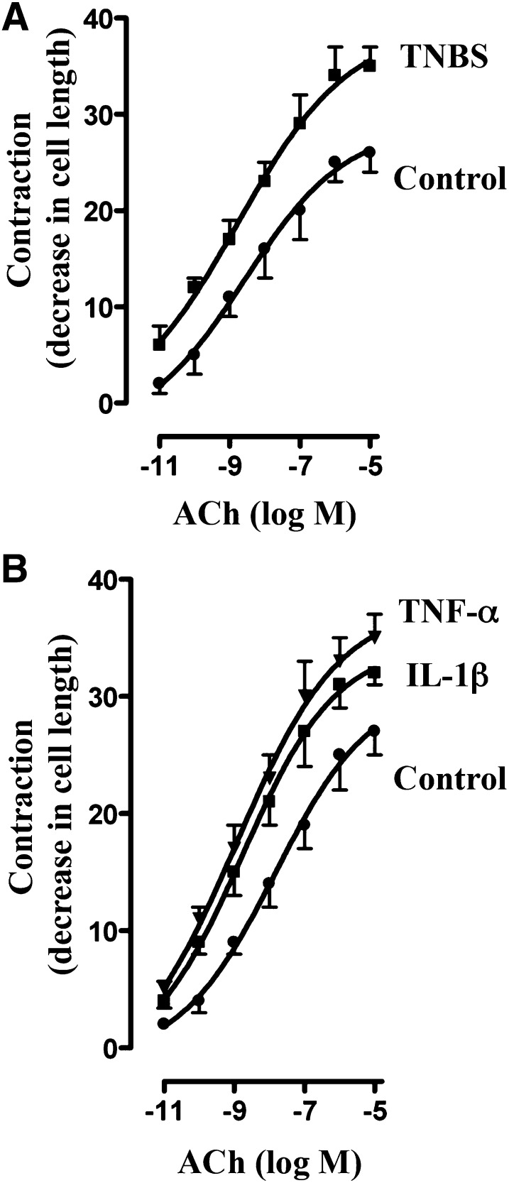
Fig. 2.
Augmentation of ACh-induced contraction by proinflammatory cytokines. Longitudinal muscle cells isolated from colon of control and TNBS-treated mice (A) or from muscle strips cultured in the absence (control) or presence of IL-1β (10 ng/ml) or TNF-α (1 nM) for 24 hours (B) were treated with different concentrations of ACh for 30 seconds to measure initial Ca2+-dependent contraction. Muscle contraction was measured by scanning micrometry and expressed as percentage of decrease in cell length before ACh treatment. Basal cell lengths were not significantly different between control and TNBS-treated mice or between control and cytokine-treated muscle strips (115 ± 4 µm to 121 ± 6 µm). Values are means ± S.E.M. of six experiments.

