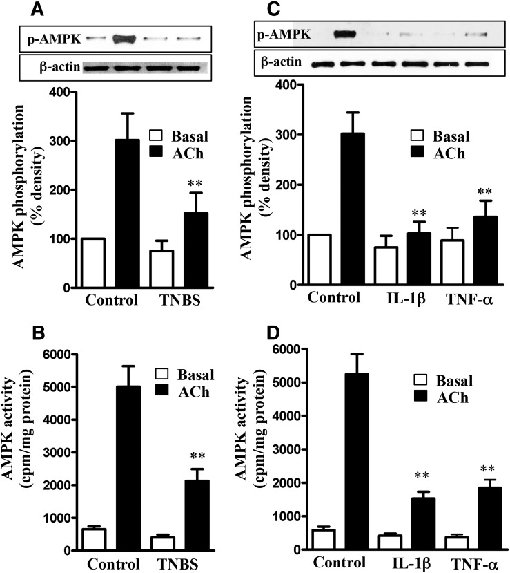
Fig. 4.
Suppression of ACh-induced AMPK phosphorylation at Thr172 and AMPK activity by proinflammatory cytokines. Longitudinal muscle cells isolated from colon of control and TNBS-treated mice (A and B) or from muscle strips cultured in the absence (control) or presence of IL-1β (10 ng/ml) or TNF-α (1 nM) for 24 hours (C and D) were treated with 1 µM of ACh for 30 seconds to measure initial Ca2+/calmodulin-dependent AMPK phosphorylation (A and C) and AMPK activity (B and D). AMPK phosphorylation at Thr172 was measured by immunoblot using phospho-specific antibody, and AMPK activity was measured by immunokinase assay. Basal AMPK activity was not significantly different between control and TNBS-treated mice or between control and cytokine-treated muscle strips. Values are means ± S.E.M. of five experiments. **P < 0.01, significantly different from control response to ACh.

