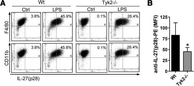
Figure 2. Lower frequencies of IL-27(p28)+F4/80+CD11b+ macrophages derived from Tyk2−/− mice compared with WT mice.

(A) Flow cytometry for intracellular IL-27(p28) and surface markers F4/80 and CD11b in TLR4-activated BMDM derived from WT mice or Tyk2−/− mice; LPS (1 μg/ml), 2 μM monensin, 12 h. Ctrl, BMDM treated with 2 μM monensin but without LPS. (B) Mean fluorescence intensity (MFI) of anti-IL-27(p28) PE-labeled antibody staining in LPS-activated BMDM from Tyk2−/− mice or WT mice. Data are shown as representatives of three independent experiments (A) or were pooled from three independent experiments (B), each done in duplicates. Student's two-tailed t-test; mean ± sem; *P < 0.05.
