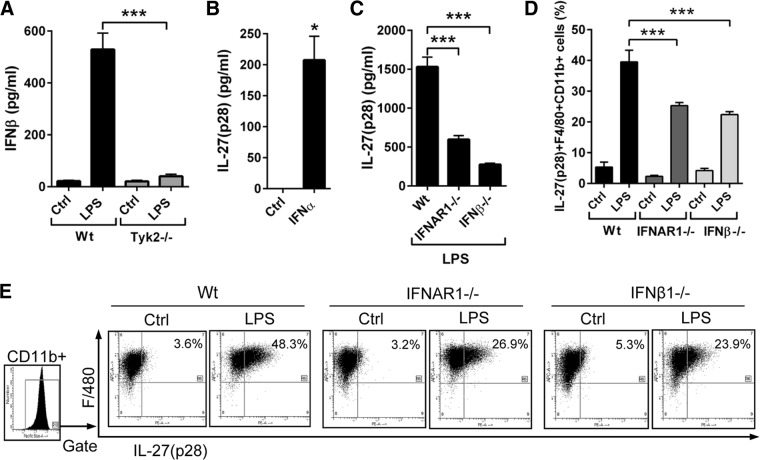
Figure 4. Tyk2 activates the endogenous production of type I IFNs for enhancing the release of IL-27(p28).
(A) Detection of IFN-β secretion by LPS-activated macrophages from WT mice or Tyk2−/− mice; Ctrl, unstimulated control macrophages; 8 h; ELISA. (B) Induction of IL-27(p28) release from BMDM (WT) after incubation with IFN-α (1000 U/ml); 8 h; ELISA. (C) IL-27(p28) release of BMDM from WT mice, IFNAR1−/− mice, or IFN-β−/− mice after LPS; 8 h. (D) Flow cytometry of IL-27(p28)+F4/80+CD11b+ BMDM from WT mice, IFNAR1−/− mice, or IFN-β−/− mice after LPS; 12 h; monensin (2 μM). Numbers indicate the percentage of IL-27(p28)+F4/80+CD11b+ cells. (E) Representative dot-plot images of the experiments described in D; all dot-plots were gated on CD11b+ cells. Data are pooled from three independent experiments (A–C) or done with n = 4 mice/group (D and E). One-way ANOVA (A, C, and D) or Student's two-tailed t-test (B); mean ± sem; *P < 0.05; ***P < 0.001.

