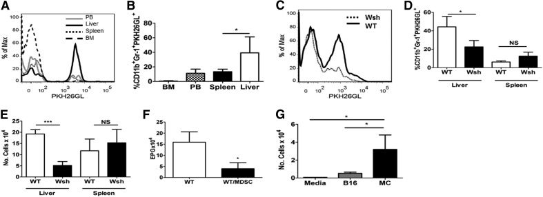
Figure 1. MDSCs preferentially migrate to the liver in a MC-dependent manner.
(A) WT mice given labeled MDSCs, analyzed 18 h later. MDSC in liver, peripheral blood (PB), spleen, and bone marrow (BM). Data compiled in B. (C) Wsh or WT was given labeled MDSCs, infected with Nb, and examined for MDSC staining on Day 7. Cell percentage of PKH26GL+ cells out of total Gr1+CD11b+cells (D) and number (E) from C. (F) Eggs/gm feces (EPG) determined on Day 7. MDSCs given Days −1, 2, and 5. (G) Four-hour MDSC migration in response to B16 melanoma, MCs, or media alone. *P < 0.05; ***P < 0.0005. Mean ± sd; n ≥ 5/group.

