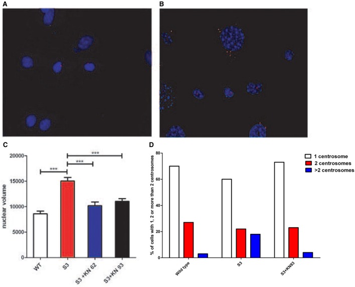
Figure 2.
(A) Centrosomes of cultured WT fibroblasts were stained with an antibody raised against γ-tubulin (in red). (B) Centrosomes of cultured S3 transgenic fibroblasts were stained with an antibody raised against γ-tubulin (in red). An abnormal number of centrosomes could be observed. (C) Nuclear volumes of S3 fibroblasts and S3 fibroblasts treated either with KN93 or KN62 were evaluated after DAPI staining, using ImageJ software. (D) Fibroblasts from WT, S3, and S3 treated with KN93 were cultured and the percentages of cells with one centrosome (white), two centrosomes (red), and more than two (blue) were determined. This analysis has been repeated three times. ***p-value < 0.001.

