
Fig. 1.
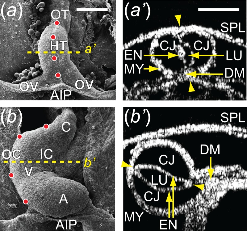
Cardiac c-looping of chick embryo. ((a) and (b)) SEM images of embryonic chick hearts during c-looping (ventral view). The originally straight HT at HH10 (a) bends ventrally and rotates rightward, transforming into a c-shaped tube at HH12 (b). To help visualize rotation, artificial labels (dots) along the ventral midline of the HT at HH10 move to the outer curvature of the HH12 heart. The splanchnopleure membrane was removed to reveal the HT. ((a′) and (b′)) Rotation of the HT is shown by the orientation of the elliptical lumen (arrowheads) in OCT cross sections taken midway along the length of the HT (dashed lines in (a) and (b)). A = atrium, AIP = anterior intestinal portal, C = conotruncus, CJ = cardiac jelly, DM = dorsal mesocardium, EN = endocardium, IC = inner curvature, LU = lumen, HT = heart tube, MY = myocardium, SPL = splanchnopleure, OC = outer curvature, OT = outflow tract, OV = omphalomesenteric veins, V = ventricle. Scale bars: 200 μm.