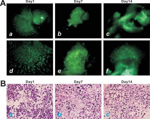
Figure 3.
Decellularized liver scaffolds support iHPC proliferation in 3-D culture in vitro. (A) Subconfluent iHPCs infected with AdGFP were perfused into the decellularized liver scaffolds (approximately 10^e7 cells/scaffold). GFP signal was detected at day 1 (a & d), day 7 (b & e), and day 14 (c & f). Panels a-c, lower magnification; Panels d-f, higher magnification. (B) H & E staining of the decullarized liver scaffolds seeded with iHPCs. The iHPC-seeded scaffolds were harvested at day 1 (a), day 7 (b), and day 14 (c), and frozen sectioned for H & E staining. Representative results are shown.

