Figure 5.
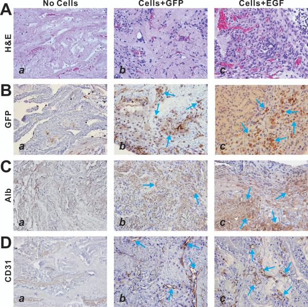
EGF-stimulated iHPC cells can proliferate and differentiate in the decullarized liver scaffolds in vivo. The renal capsule grafting samples were retrieved at 10 days post implantation and subjected to frozen sectioning. The sections were subjected to H & E staining (A), anti-GFP immunohistochemical staining (B), anti-albumin immunohistochemical staining (C), and anti-CD31 immunohistochemical staining (D). Control IgGs were used as negative controls (data not shown). Arrows indicate positive staining cells. Representative results are shown.

