
Figure 3. Differences Between Infant Apolipoprotein E ε4 Carriers and Noncarriers in Regional Gray Matter Volumes (GMVs).
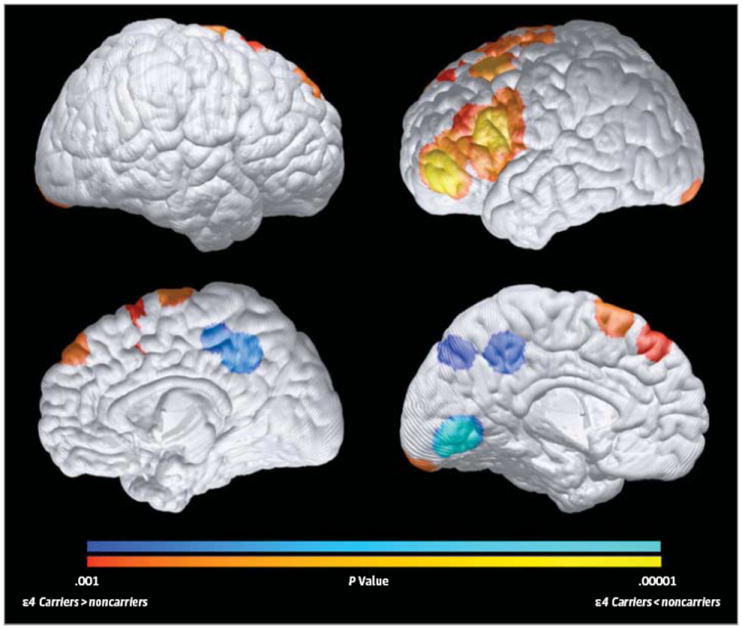
Compared with noncarriers, 6- to 22-month-old ε4 carriers had significantly reduced GMVs in the bilateral precuneus, posterior/middle cingulate, and occipitotemporal regions (as shown in blue) and in a left lateral temporal region (not shown, because it is too deep to be projected onto the cortical surface), which are preferentially affected in the later preclinical and clinical stages of Alzheimer disease, and significantly greater GMVs (in red) in bilateral medial and lateral frontal regions (P < .001, uncorrected for multiple comparisons). Statistical maps are projected onto the medial and lateral surfaces of a spatially standardized 12-month-old infant’s brain. The magnitude and atlas locations of maximally significant differences in regional GMV are shown in Table 1.