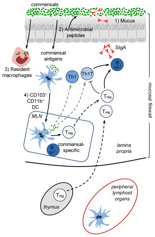
Figure 1. The mucosal firewall.
1) The mucus represents the primary barrier limiting contact between the microbiota and host tissue and preventing microbial translocation. 2) Epithelial cells produce antimicrobial peptides that also play a significant role in limiting exposure to the commensal microbiota. 3) Translocating commensals are rapidly eliminated by tissue resident macrophages. 4) Commensals can also be captured by CD103+ CD11b+ DCs that traffic to the mLN from the lamina propria but do not penetrate further. Presentation of commensal antigens by these DCs leads to the differentiation of commensal specific regulatory cells (Treg) Th17 cells and IgA producing B cells. Commensal specific lymphocytes traffic to the lamina propria and Peyer’s Patches. In the Peyer’s patches Treg can further promote class switching and IgA generation against commensals. The combination of the epithelial barrier, mucus layer, IgA and DCs and T cells comprises the ‘mucosal firewall’, which limits the passage and exposure of commensals to the Gut-Associated Lymphoid tissue preventing untoward activation and pathology.

