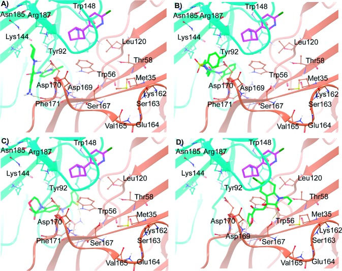
Figure 4.
Molecular interactions of the compounds 1, 2, 4, and 9, represented by A, B, C, and D, respectively, predicted by Glide XP docking. The monomer chains (ribbon representation) and carbon atoms from each chain are differentially colored (α4 = cyan, and β2 = light orange). Epibatidine is shown as a stick representation with purple carbon, whereas the docked compounds are represented as stick green carbon. Relevant amino acid residues in the binding site are shown in ball and stick representation.

