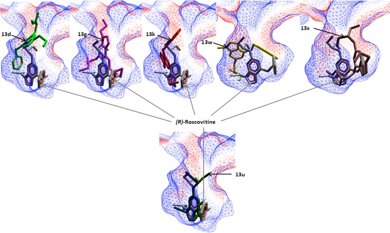
Figure 2.
Docking of 13d, 13g, 13k, 13w, 13x, and 13u to the cdk2/roscovitine complex. The electrostatic interaction surface at the binding site region is displayed and colored red for negative charge and blue for positive charge. Docking simulations were performed using Molegro Virtual Docker, taking into account side chain flexibility for all residues in the binding region.

