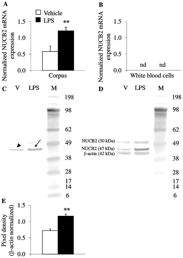
Fig. 2.

LPS increases gastric corpus NUCB2 mRNA and protein concentration, whereas NUCB2 mRNA is not detectable in white blood cells. Ad libitum fed rats were injected ip with LPS (100 μg/kg body weight) or vehicle (pyrogen-free saline) and trunk blood and stomach were collected at 2 h post injection. (A and B) Gastric corpus and white blood cell mRNA were isolated and assessed for NUCB2, GAPDH and β-actin. After injection of LPS, NUCB2 mRNA expression (normalized for housekeeping genes, GAPDH and β-actin) was increased in the gastric corpus compared to vehicle (A) but was not detectable in white blood cells (B). (C–E) Equal amounts of gastric corpus protein were loaded and NUCB2/nesfatin-1 concentrations assessed using Western blot followed by semi-quantitative analysis. Lane 1 contains corpus after vehicle injection, lane 2 corpus proteins after LPS and lane 3 the molecular weight standards (C). The Western blot shows full length NUCB2 with (∼50 kDa) or without signal sequence (∼ 47 kDa) but not mature nesfatin-1 (∼10 kDa). Injection of LPS increased NUCB2 (arrow) compared to vehicle (arrowhead, C). Re-staining of the Western blot with ß-actin (42 kDa) demonstrated equal gastric corpus mucosal protein concentration (D). Quantification of NUCB2 stomach protein expression is shown in (E). Each bar represents the mean ± SEM of 5 rats/group. **p < 0.01 vs. vehicle.
