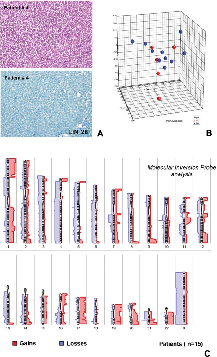
Fig. 1.
Principal component analysis and virtual karyotype results from the molecular inversion probe analysis of supratentorial CNS-PNET cases. The cases analyzed in this study commonly showed CNS-PNET histology with undifferentiated, small-cell cytology. (A, upper part: hematoxylin and eosin). All tumors included in the study were LIN28A negative (A, lower part: immunostaining with LIN28A antibody). The principal component analysis showed molecular heterogeneity of the 15 supratentorial CNS-PNETs examined with MIP. (B) No molecular subgroups were identifiable according to patient age (red points represent patients <4 years of age; blue points represent patients >4 years). The virtual karyotype (C) showed frequent gains of 1q, 17, 19, 20, and 22 as well as losses of 1p (in particular 1p33-1p21), 3, 4, 5, and 13. No case presented focal chromosome 19q13.41 amplification. Gains were indicated by the bars (red) on the right side of each chromosome, and losses (blue) are shown on the left side. Thicker bars indicate areas of recurrent change.

