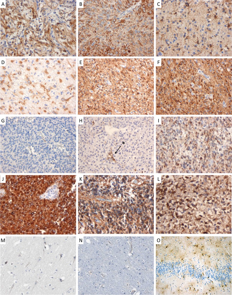
Fig. 2.
PBXIP1 immunohistochemistry on paraffin-embedded sections. Representative images of cytoplasmic PBXIP1 staining in WHO grade I pilocytic astrocytoma (A and B); WHO grade II diffuse astrocytoma (C and D); WHO grade III anaplastic astrocytoma (E and F); WHO grade III anaplastic oligodendroglioma (G and H; arrow and asterisk indicate reactive astrocytes); WHO grade IV glioblastoma multiforme (I and J); WHO grade III anaplastic ependymoma (K and L); non-malignant cerebral cortex (M and N) and reactive astrogliosis (O).

